Introduction

        Haemolytic disease of the newborn, also known as haemolytic disease of the foetus and newborn, HDN, HDFN, or erythroblastosis fetalis, is an alloimmune condition that develops in a foetus, when the IgG molecules produced by the mother pass through the placenta. During pregnancy, IgG antibodies are actively transported across the placenta. If the foetus has the RBC antigen corresponding to the mother’s antibody, the antigen-antibody interaction can result in the destruction of the foetal RBCs.


      Among these antibodies are some which attack the red blood cells in the foetal circulation; the red cells are broken down and the foetus can develop reticulocytosis and anaemia. This foetal disease ranges from mild to very severe, and foetal death from heart failure (hydrops fetalis) can occur. When the disease is moderate or severe, many erythroblasts are present in the foetal blood and so these forms of the disease can be called erythroblastosis fetalis. 

Signs & Symptoms

The following are the most common symptoms of haemolytic disease of the newborn. However, each infant may experience symptoms differently. During pregnancy symptoms may include:

Before birth

  • With amniocentesis, the amniotic fluid may have a yellow colouring and contain bilirubin.

  • Ultrasound of the foetus shows enlarged liver, spleen, or heart and fluid build up in the foetus' abdomen.

After birth

Symptoms may include:

  • As the red blood cells break down, bilirubin is formed. Infants unable to get rid of the bilirubin. Builds up of bilirubin in the blood (hyperbilirubinemia) and other tissues and fluids of the infant's body resulting in jaundice.

  • Jaundice or yellow colouring of amniotic fluid, umbilical cord, skin, and eyes may be present. The  infant  may not look yellow immediately after birth, but jaundice can develop quickly, usually within 24 to 36 hours. Beside that, maternal antibodies destroy foetal red blood cells that results in anaemia. Anaemia limits the ability of the blood to carry oxygen to the  infant's organs and tissues and lead to breathing difficulties to the  infant.
Jaundice


  • Infant's responds to the haemolysis by trying to make more red blood cells very quickly in the bone marrow and the liver and spleen organs and result in enlargement of these organ and known as hepatospleenomegaly. Furthermore, new red blood cells released prematurely from bone marrow and are unable to do the work of mature red blood cells are remove to the spleen also lead to spleenomegaly


Complication

Kernicterus 


Kernicterus also will occur when unconjugated bilirubin can reach levels toxic to the infant’s brain (generally 18mg/dL). Symptoms of non-life threatening kernicterus can range from deafness to severe mental retardation.


Kernicterus


         Hydrops Fetalis


Beside that, Hydrops Fetalis will occur as the baby's organs are unable to handle the anaemia. The heart begins to fail and large amounts of fluid build up in the baby's tissues and organs. In severe forms this can include petechiae and purpura. The infant may be stillborn or die shortly after birth.

Hydrop fetalis

Pathogenesis

Before birth


  • Antigen-antibody interaction occurs.
  • Antibody coated RBCs are removed from foetal circulation by macrophages of the spleen and liver.
  • Results: anaemia
  • In response to anaemia, foetal bone marrow and other haematopoietic tissues in the spleen and liver increase the amount of RBCs produced.
  • Foetal anemia may range from mild to severe depending on the amount of foetal RBCs destroyed and the capacity of the erythropoiesis to compensate.
  • If severe, the foetus may develop hydrops fetalis.
  • As RBCs haemolyze, haemoglobin is released and metabolized to indirect bilirubin
  • Indirect bilurubin is transported across placenta and conjugated to direct bilirubin in the mother’s liver.
  • Conjugated bilirubin is then excreted by the mother and does not cause clinical disease in the fetus.


       After birth


  • The rate of RBCs destruction after birth decreases because there is no additional antibody entering the infant’s circulation through placenta.
  • After the infant is born, its liver is unable to conjugate bilirubin efficiently because of a deficiency in the enzyme, glucuronyl transferase.
  • Result: accumulation of metabolic by products of RBCs destruction can become severe problem for the newborn.
  • The unconjugated bilirubin can reach levels toxic to infant’s brain (generally 18mg/dL), cause kernicterus or permanent damage to parts of the brain. 

Aetiology

  • Antibody of RBCs potentially cause HDN is antibody of IgG class that possessed by the mother and the antibody is known could cause various degree of complications to the foetus if the foetus has RBCs containing the corresponding antigen. 
  • IgG class antibody is important in HDN because their ability to cross placenta and are actively transported cross the placenta.

  • Other factors that determine the degree and the incident of HDN are (Quinley, 1998):
                         - Development degree of RBCs antigen
                         - Specificity, avidity and class of the antibody
                         - Maternal antibody titre

  • There are three major aetiology of HDN, which are:
  1. Rh HDN
  2. ABO HDN
  3. Other alloantibodies

  Rh HDN

  • Rh HDN is the most severe form of HDN among the three aetiology of HDN and sometimes fatal. 
  • Rh HDN happens when mother, who is D negative, bearing D positive foetus. 
  • D positivity of the foetus is due to genetically inheritance of D from father.  
  • If the father is heterozygous for the RHD deletion, there is a 50% chance of the foetus being D-negative. 
  • If the father is homozygous for the RHD gene, the foetus will definitely inherit the D antigen. 
  • D- mother who lack a functional RHD gene do not produce the D antigen, and may be immunized by D-positive foetal blood.

  • Anti-D is the commonest antibody in Rh system that cause HDN and the complication vary from moderate to severe. 
  • Second most common is anti-E that can cause mild HDN complication. 
  • Anti-c can range from a mild to severe complication. 
  • Other two antibodies of Rhesus group (anti-e and anti-C) are rare to be reported. 
  • Combination of Rhesus antibodies can be severe.

  • Sensitization usually occurs very late in Rh-positive pregnancy, hence first Rh-positive child is not affected but the second Rh-positive child will be equally or severely affected. 
  • However, sensitization at first Rh-positive pregnancy can occur because foetus express Rh antigen on their RBCs membrane after 4 weeks of gestation. 
  • Anti-D developed usually not detected before week 35 of gestation and the antibody titre is usually low, therefore, first Rh-positive child seldom shows clinically significant HDN.

  • Sensitization can occur through of fetomaternal haemorrhage (delivery, placental membrane rupture, trauma to abdomen, amniocentesis, abortion etc.) and previous transfusion of incompatible Rhesus blood group. 
  • Degree of sensitization varies according to individual (example: immune response) and level of exposure to Rh-positive RBCs.

ABO HDN

  • ABO HDN is the most common form of HDN. 
  • Anti-A and anti-B in person of A and B blood group are mostly of IgM class and cause no isoimmunisation to occur. 
  • In the other hand, anti-A and anti-B in O blood group person are partially of IgG class. 
  • Therefore, when O blood group mother is gestating A or B blood group foetus, there is a possibility of HDN to occur. 
  • ABO HDN can occur in the first pregnancy due to unnecessary need for sensitization since ABO antibodies is readily present in the blood system (natural occurring antibodies). 

  • ABO HDN has mild complication and RBCs destruction is usually short-live (jaundice and anaemia only evident within 12 to 48 hours after birth)
  • This mild complication occurs is may be due to (Quinley, 1998):
    • Smaller number of A and B antigenic site on foetal RBC membrane
    • Foetal RBCs are less developed at birth, hence less RBC destruction by maternal antibodies
    • Presence of A and B antigen on other than RBCs surfaces such as tissues and secretions cause the ABO antibodies fractionate to combine with the antigens, therefore less antibodies that cross placenta combine to foetal RBCs 

Laboratory Diagnosis

a. Detection of HDN can be done through prenatal and postnatal test.
b. Prenatal test is performed to identify those women at risk of having babies affected with HDN. Specimens used are clotted blood sample from both mother and father.
c. Similar to prenatal test, postnatal also used clotted blood sample, but taken from mother and infant.
d. Cord blood sample is taken through needle aspiration, and should be delivered and refrigerated for minimum of 7 days.
e. The severity of HDN can be access through in vitro assays, that is:
                  • Antibody titration
                  • Quantifying anti-D
                  • Maternal history
                  • IgG subclass distribution


Figure 1: Summary of diagnosis test flow.


Prenatal Laboratory Test


  • ABO and rhesus (Rh) typing
                    
                   ABO and Rh typing is done both on father and mother sample. In addition, weak D is also done to confirm the true Rh-negative, if the result shows Rh-negative. In Rh-HDN, paternal Rh phenotyping can be very helpful in predicting future pregnancies outcome. It’s not a problem if both of the parents are rhesus-positive. But, rhesus-negative mother and the rhesus-positive father who are getting a baby, there are possibilities for the baby to suffered from HDN. Other HDN-type used this test in determining whether the father’s RBC is homozygous or heterozygous for the gene producing immunizing antigen. It should be borne by testing with anti-D, -C, -c, -E and -e, in determining the infant’s degree of risk of exposure. This should be done in all cases involving clinically significant antibody.  At about 12 weeks of gestation, Rh-negative women should be tested for anti-D. Usually, Rh typing of maternal sample is done especially in second or subsequent pregnancy. Titration of anti-D is done to find out the strength and rising trend of titre – done by two-weekly/monthly anti-D titre determination


  • Antibody screening


                   Antibody screening test is done after ABO and Rh typing to detect if any unexpected antibodies present within mother’s blood. It is repeated at week 28 prior to Rhogam (RhIg) administration in Rh-negative women. Once detected, antibody identification is done in order to identify the antibody present, either significant or insignificant antibodies. The problem comes when the unexpected antibodies are the significant one. Titration is done if the antibodies detected are the significant ones.


  • Titration
  
                  Because other factors vary, such as the Rh-antibody–binding constant, the amount of D antigen on the RBC membrane, and the ability of the fetus to maintain reasonable circulating RBC hemoglobin without compromising hepatocellular function and umbilical portal venous circulation, Rh-antibody titrations predict only the fetus at risk of suffering from erythroblastosis erythromatosis. Beside, titration can be done for two purposes: 1) distinguishing between mildly affected infants with others, and 2) detecting increase antibody level on serial testing. Due to its sensitiveness, many laboratories used indirect antiglobulin test compare to albumin test. The indirect antiglobulin titer that puts the fetus at risk may be 1:32 to 1:64 but must be determined individually for each laboratory. Regular antibody titrations must be carried out during pregnancy because the antibody titer is the basis for selecting the mother and her fetus whose are at risk and require further investigation. Methods for titration vary from one laboratory to another. The first sample is freeze if subsequent titer is requested, so that a comparison between first titer results with second titer can be done. After run both titers in parallel, the end point is compare and if it shows rising of two-fold or greater, it is consider clinically significant. A fourfold increase in titer is significant and implies that that the fetus is Rh positive and is potentially affected. A more sensitive test is done, that is amniocentesis.


  • Amniocentesis                  


Figure 2:   Diagram flow of amniocentesis done 
in pregnant women with antibody.
Amniocentesis is used for detection of bilirubin level, fetal lung maturity profile, biophysical profiles and middle cerebral artery peak systolic velocity. It allows spectrophotometric measurement of deviation in OD at 450nm due to bilirubin level in amniotic fluid, which reflects fetal RBC hemolysis. Different degrees of change are measured or interpret differently, because the normal level of bilirubin varies with gestational age. Amniocentesis procedure should be carried out only after careful ultrasound placental localization. If amniocentesis must be carried out without the use of ultrasound, a suprapubic approach may be less likely to encounter the placenta. Nevertheless, amniocentesis carried out without ultrasound guidance is associated with a 10% to 11% risk of fetomaternal TPH and increased severity of Rh disease. This test is more reliably accurate to perform in third trimester pregnancy rather than second trimester. Usually, amniocentesis performed on women with alloantibody or has an antibody titers at or greater than critical level. Figure 2 below stated the diagram flow of amniocentesis done in pregnant women with clinically significant antibody.


Figure 3:  Liley graph
             Figure 3 shows Liley graph plotted in reading amniocentesis result. Basically, this graph is divided into 3 parts: 1) lower zone - indicates either no disease or only a mildly affected fetus. 10% require exchange transfusion; 2) mid zone - indicate a moderately affected fetus, which is a candidate for an early delivery & possible exchange transfusion; and 3) upperzone - fetus require immediate attention as severed disease is predicted.

                    For those who can't imagine how the amniocentesis is done, lets look at the video to make things clearer:


  • Percutaneous umbilical cord blood sampling (PUBS)


                    With the development of sophisticated ultrasound equipment and the availability of perinatologists skilled in its use, percutaneous fetal umbilical blood sampling became feasible in the mid-1980s. This procedure allows the direct measurement of all blood parameters that can be measured after birth (hemoglobin, hematocrit, blood groups, direct antiglobulin testing, serum bilirubin levels, platelet and leukocyte counts, serum protein levels, and fetal blood gases). In addition, this technique also allows for direct fetal transfusion. Fetal blood sampling is by far the most accurate means of determining the degree of severity of fetal hemolytic disease (in the absence of hydrops) and the need for fetal treatment measures. Fetal blood sampling is a relatively benign procedure, carrying with it a traumatic fetal mortality rate of a fraction of 1%. Because it carries a high risk of fetomaternal TPH, its use is recommended only when serial amniotic fluid ΔOD 450 readings rise into the upper 65% to 75% level of zone 2 or when an anterior placenta cannot be avoided at amniocentesis and maternal pregnancy history or maternal alloantibody titers place the fetus at risk. Fetal blood sampling may be possible as early as 18 weeks' gestation; it usually is feasible by 20 to 21 weeks' gestation. The preferred sampling site is from the umbilical vessel (preferably the vein) at its insertion into the placenta. For this reason, the procedure is technically easier if the placenta is implanted on the anterior uterine wall. The risks of PUBS include amnionitis, premature rupture of membranes, fetal loss, puncture of the umbilical artery and umbilical cord hematoma.


Postnatal Laboratory Test

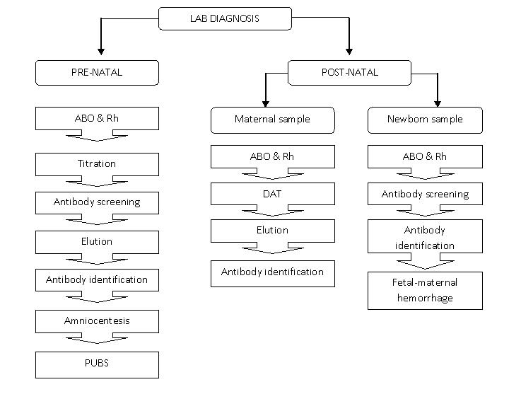
Postnatal test can be done by using both newborn and maternal blood sample.


  • ABO and Rh typing


                    As antibodies had not fully develops in newborn, only the ABO forward grouping is done on newborn sample. Rh typing is done in order to identify the baby’s Rh group. Weak D test is also performed if the baby is Rh negative. Positive Rh result obtain is not solely indicates that the newborn is Rh-positive, but it may also indicate the presence of maternal antibody that heavily coated on newborn’s RBC. Furthermore, newborns who were transfused while intrauterine often type Rh negative or weakly Rh positive. Therefore, it is impossible to get a proper Rh type without heating fetal RBC first to eluate off the antibody. ABO and Rh typing for maternal sample is done similarly to prenatal test.


  • Direct antiglobulin test (DAT) and elution.


                    DAT test should be performs on cord blood RBCs in identifying in vivo sensitization by maternal antibody. There are varies factors that contribute to the strength of DAT, which include maternal antibody strength, number of antigenic site directed by maternal antibody and total amount of blood infant receive from transfusion. Elution is done when the DAT outcome is positive. In addition, elution is also performed if the baby is symptomatic even if the DAT test shows negative result. It is unnecessary to perform elution on infant’s RBC if there is available of maternal blood and a single antibody is identified. If however, the maternal serum is negative for unexpected antibodies, so it is suggested of having ABO antibodies or an antibody to a low-frequency antigen. Diagnosing a low-frequency antigen can be aids by testing the eluate with paternal RBCs. Even though the antibody screening tests and panels give negative result, testing the maternal serum against paternal serum confirms the presence of antibody. As stated above, elution is done after getting the result of DAT.  Type of cells used to test eluate depends on antibody found on mother. Below are 2 examples of situations that shows usage of different types of elute cells.
·         Mom: anti-A, anti-B and/or anti-AB; negative antibody screening
                         Baby: A or B blood group

o   Test eluate against screening cells, A1 and B cells.
o   May performed Lui freeze-thaw or acid elution.
o   Strength of DAT varies depends on maternal antibody strength, no of antigenic sites for antigen to which maternal antibody is directed, how much blood the infants has received from transfusion.

·         Mom: positive antibody screen

o   Perform acid elution.
o   Against the same cells that react in mother ab identification panel.
o   Include reagent A1 and B cells if mother is group O, baby is A or B.


  • Antibody screening and identification


                    Antibody screening only performed on maternal sample, as the newborn did not fully develop their own antibodies. It is done similar to those procedure perform for prenatal test.


  • Fetal-maternal hemorrhage (FMH).

                    Protection of mother against the immunizing effects of Rh-positive fetal RBCs is vital, therefore it is important for the Rh-negative women to receive appropriate amount of RhIG. The half-life of the RhIG in the absence of significant mixing of fetal with maternal circulation, called fetal-maternal hemorrhage, is approximately 21 days. Two tests are commonly used to detect and quantify FMH: Rosette test and Kleihauer-Betke test.

                                                        i.            Rosette test
o   Qualitative test.
o   Used to detect FMH greater than 30mL.
o   Procedure:
                                                                                                               I.        Incubate suspension of maternal blood with anti-D
                                                                                                            II.        Fetal Rh-positive RBCs in maternal blood react with anti-D.
                                                                                                         III.         Wash unbound antibody.
                                                                                                        IV.         Add suspension of group O, Rh-positive cells.
                                                                                                          V.          Reaction between anti-D with both O Rh-positive and fetal Rh-  
                                                                positive RBCs, form agglutination in a rosette form.
                                                                                                        VI.         Examine the suspension microscopically.

o   Presence of a certain number of clumps in a defined number of microscopic fields indicates positive result.
o   Less than 30mL indicates negative result.
o   Quantitative test must be performed to estimate the number of vials of RhIg needed.

                                                      ii.            Kleihauer-Betke (KB) test.
o   Quantitative test.
o   Must be performed if positive rosette test.
o   Done to estimate the needed number of RhIG vials.
o   Principle is based on the resistance of fetal hemoglobin to acid elution.
o   The percentage of fetal cells in maternal sample can be used to calculate the volume of FMH and the appropriate number of vials of RhIG.
o   Indication:
-       Determine possible FMH in the newborn.
-       Aid in diagnosis of certain types of anemia in adults
-       Assess the magnitude of FMH
o   Calculate dosage of RhIg to be given through the percentage of fetal cells in maternal sample.

                                                    iii.            Flow cytometry
o   Similar purpose with KB test.
o   Less available compare to KB test.
o   Done by analyzing mixture of maternal sample, anti-D and fluorescein-labeled IgG by fluorescein-activated cell sorter.                           


CLINICAL FINDINGS

·         Anemia
·         Hyperbilirubinemia
·         Reticulocytosis
·         Thrombocytopenia
·         Increase nucleated RBC count
·         Leucopenia
·         Hypoalbuminemia
·         Smear: polychromasia, anisocytosis, no spherocytes.
·         Rh negative blood type
·         Positive DAT

Treatment

Once HDN is diagnosed, treatment may be needed.  Treatment for HDN can be divided to before delivery and after delivery for Rh HDN and ABO HDN.

Rh HDN


  • If HDN is detected during pregnancy (before delivery) that is Rh HDN cases,
  • it will be treated using intrauterine transfusion (IUT). Intrauterine transfusion is done if the fetus if affected severely, transfusions will be done every 1 to 4 weeks until the fetus is mature enough to be delivered safely. Amniocentesis may be done to determine the maturity of the fetus's lungs before delivery is scheduled. It is given to the fetus to prevent hydrops fetalis and fetal death. This method can be done as early as 17 weeks, although preferable to wait until 20 weeks. After multiple IUTs, most of the baby’s blood will be D negative donor blood, therefore, the Direct Antiglobulin test will be negative, but the Indirect Antiglobulin Test will be positive. After IUTs, the cord bilirubin is not an accurate indicator of rate of hemolysis or of the likelihood of the need for post-natal exchange transfusion.

  • IUTs procedure will be firstly done by sedating the mother, and an ultrasound image is obtained to determine the position of the fetus and placenta. After the mother's abdomen is cleaned with an antiseptic solution, she is given a local anesthetic injection to numb the abdominal area where the transfusion needle will be inserted. Medication may be given to the fetus to temporarily stop fetal movement. Ultrasound is used to guide the needle through the mother's abdomen into the fetus's abdomen or an umbilical cord vein. A compatible blood type (usually type O, Rh-negative) is delivered into the fetus's abdominal cavity or into an umbilical cord blood vessel. The mother is usually given antibiotics to prevent infection. She may also be given tocolytic medication to prevent labor from beginning, though this is unusual. The risk of these procedures is now largely dependent on the prior condition of the fetus and the gestational age at which transfusion is commenced.

Intrauterine Transfusion


Prevention

  1. ABO HDN, no way to prevent or predict.
  2. For Rh HDN there are ways to prevent the occurrence of Rh HDN.
  
  • Fortunately, HDN is a very preventable disease. Because of the advances in prenatal care, nearly all women with Rh negative blood are identified in early pregnancy by blood testing. If a mother is Rh negative and has not been sensitized, she is usually given a drug called Rh immunoglobulin (RhIg), also known as RhoGAM. This is a specially developed blood product that can prevent an Rh negative mother's antibodies from being able to react to Rh positive cells. Many women are given RhoGAM around the 28th week of pregnancy. After the baby is born, a woman should receive a second dose of the drug within 72 hours.


Rhogam

 
Haemolytic Disease of the Newborn (HDN) © 2012 | Designed by Canvas Art, in collaboration with Business Listings , Radio stations and Corporate Office Headquarters